Garden City Eye Hospital
icon-clock-medic.png
Mon - Friday 10:00am-8:00pm

Sunday - Only on prior Appointments

National Accreditation Board
for Hospitals & Healthcare Providers
Monday - Friday 08:00-19:00

Saturday and Sunday - CLOSED

+62 810 2633 8711

csmedison@example.com

1428 Callison Laney Buoy

Building 201/VRY Virginia, VA 22902

Vitreo-Retinal & Uvea

Vitreo-Retinal & Uvea

Retinal diseases vary widely, but most of them cause visual symptoms. Retinal diseases can affect any part of your retina, a thin layer of tissue on the inside back wall of your eye.

The retina contains millions of light-sensitive cells (rods and cones) and other nerve cells that receive and organize visual information. Your retina sends this information to your brain through your optic nerve, enabling you to see.

Treatment is available for some retinal diseases. Depending on your condition, treatment goals may be to stop or slow the disease and preserve, improve or restore your vision. Untreated, some retinal diseases can cause severe vision loss or blindness.

SYMPTOMS

Many retinal diseases share some common signs and symptoms. These may include:

  • Seeing floating specks or cobwebs
  • Blurred or distorted (straight lines look wavy) vision
  • Defects in the side vision
  • Lost vision

WHEN TO SEE A DOCTOR ?

 
It’s important to pay attention to any changes in your vision and find care quickly. Seek immediate medical attention if you suddenly have floaters, flashes or reduced vision. These are warning signs of potentially serious retinal disease.

TYPES

Common retinal diseases and conditions include :
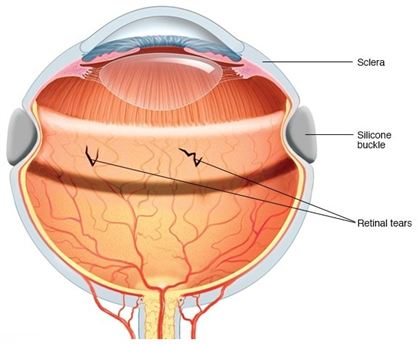
  • Retinal tear: A retinal tear occurs when the clear, gel-like substance in the center of your eye (vitreous) shrinks and tugs on the thin layer of tissue lining the back of your eye (retina) with enough traction to cause a break in the tissue. It’s often accompanied by the sudden onset of symptoms such as floaters and flashing lights.
  • Retinal detachment: A retinal detachment is defined by the presence of fluid under the retina. This usually occurs when fluid passes through a retinal tear, causing the retina to lift away from the underlying tissue layers.
  • Diabetic retinopathy: If you have diabetes, the tiny blood vessels (capillaries) in the back of your eye can deteriorate and leak fluid into and under the retina. This causes the retina to swell, which may blur or distort your vision. Or you may develop new, abnormal capillaries that break and bleed. This also worsens your vision.
  • Epiretinal membrane: Epiretinal membrane is a delicate tissue-like scar or membrane that looks like crinkled cellophane lying on top of the retina. This membrane pulls up on the retina, which distorts your vision. Objects may appear blurred or crooked.
  • Macular hole: A macular hole is a small defect in the center of the retina at the back of your eye (macula). The hole may develop from abnormal traction between the retina and the vitreous, or it may follow an injury to the eye.
  • Macular degeneration: In macular degeneration, the center of your retina begins to deteriorate. This causes symptoms such as blurred central vision or a blind spot in the center of the visual field. There are two types — wet macular degeneration and dry macular degeneration. Many people will first have the dry form, which can progress to the wet form in one or both eyes.
  • Retinitis pigmentosa: Retinitis pigmentosa is a degenerative disease that affects the retina.
  •  
  •  
  •  
  •  

TREATMENT

Scleral buckle
The main goals of treatment are to stop or slow disease progression and preserve, improve or restore your vision. In many cases, damage that has already occurred can’t be reversed, making early detection important. Your doctor will work with you to determine the best treatment. Treatment of retinal disease may be complex and sometimes urgent.
Treatment Options include:
  • Using a laser: Laser surgery can repair a retinal tear or hole. Your surgeon uses a laser to heat small pinpoints on the retina. This creates scarring that usually binds (welds) the retina to the underlying tissue. Immediate laser treatment of a new retinal tear can decrease the chance of it causing a retinal detachment.
  • Shrinking abnormal blood vessels: 
    Your doctor may use a technique called scatter laser photocoagulation to shrink abnormal new blood vessels that are bleeding or threatening to bleed into the eye. This treatment may help people with diabetic retinopathy. Extensive use of this treatment may cause the loss of some side (peripheral) or night vision.
  • Freezing: 
    In this process, called cryopexy (KRY-o-pek-see), your surgeon applies a freezing probe to the external wall of the eye to treat a retinal tear. Intense cold reaches the inside of the eye and freezes the retina. The treated area will later scar and secure the retina to the eye wall.
  • Injecting air or gas into your eye: 
    This technique, called pneumatic retinopexy (RET-ih-no-pek-see), is used to help repair certain types of retinal detachment. It can be used in combination with cryopexy or laser photocoagulation.
  • Indenting the surface of your eye: 
    This surgery, called scleral (SKLEER-ul) buckling, is used to repair a retinal detachment. Your surgeon sews a small piece of silicone material to the outside eye surface (sclera). This indents the sclera and relieves some of the force caused by the vitreous tugging on the retina. This technique may be used with other treatments.
  • Evacuating and replacing the fluid in the eye: 
    In this procedure, called vitrectomy (vih-TREK-tuh-me), your surgeon removes the gel-like fluid that fills the inside of your eye (vitreous). He or she then injects air, gas or liquid into the space.
  • Vitrectomy
     may be used if bleeding or inflammation clouds the vitreous and obstructs the surgeon’s view of the retina. This technique may be part of the treatment for people with a retinal tear, diabetic retinopathy, a macular hole, an infection, eye trauma or a retinal detachment.
  • Injecting medicine into the eye: 
    Your doctor may suggest injecting medication into the vitreous in the eye. This technique may be effective in treating people with wet macular degeneration, diabetic retinopathy or broken blood vessels within the eye.
  • Implanting a retinal prosthesis: 
    People who have severe vision loss or blindness owing to retinal disease may be a candidate for surgery. One surgical option not widely available is called retinal prosthesis.